 Introducing our BRAND NEW brePRINT.
  10 Min Read
 Kelly Mills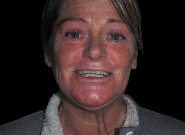
Patient expectations are rising. It is no longer sufficient to be able to offer implants as an alternative to dentures or conventional bridges. Nowadays, people with gaps or failing teeth want an option that satisfies their need for speed, function and aesthetics from day one of treatment. This is none more apparent than with edentulous patients who wish to walk out of the practice on the day of surgery with a full set of fixed, temporary teeth.
Procedures such as immediate implant placement, immediate loading and ‘teeth-in-a-day’, respond to such growing demand. As dentists, we are constantly seeking ways to provide implant treatment more precisely and efficiently.
We strive to optimise chair time, reduce the number of surgery visits, minimise the risk of errors and complications and, above all, enhance the entire patient experience.
Along with mastering the art of teeth-in-a-day procedures and careful selection of the implants and associated systems, photogrammetry comes to the implantologist’s aid. The technology offers a transfer system capable of accurately calculating the position and orientation of implant interfaces. Extraoral scans enable the clinician to capture precise data about implants with photographs of the patient’s dentition and other structures, to create a very exact three-dimensional model of the mouth. Integration of photogrammetry into the workflow for full-arch rehabilitation avoids the need for a verification jig, which is traditionally used to check that a master model is correct before fabrication of the framework.
Case presentation and examination
The following case describes full-arch upper and lower implant treatment with bredent medical copaSKY implants, multi-unit abutments and abutment screws, and bredent medical SKY fast & fixed same-day-teeth therapy. Treatment planning was also aided with an iTero intraoral scanner and Tupel 3D photogrammetry extraoral scanner, for which bredent medical provides scan bodies. The case was supported by London-based dental laboratory, Biterite, and lead technician, Michael Joseph.
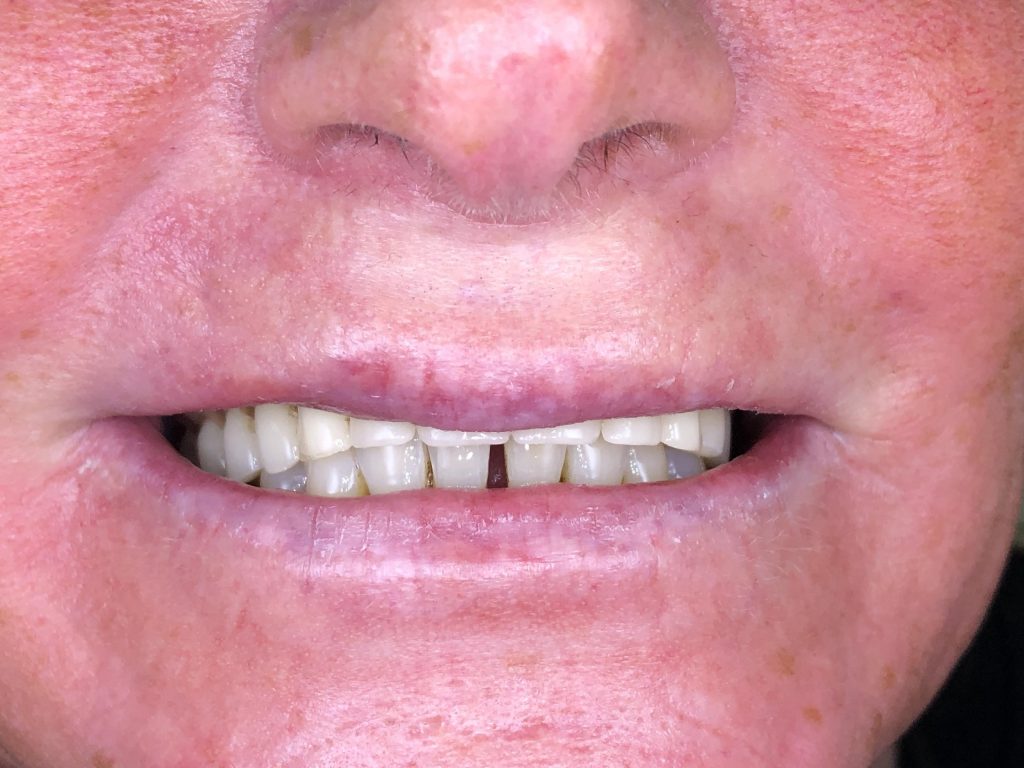
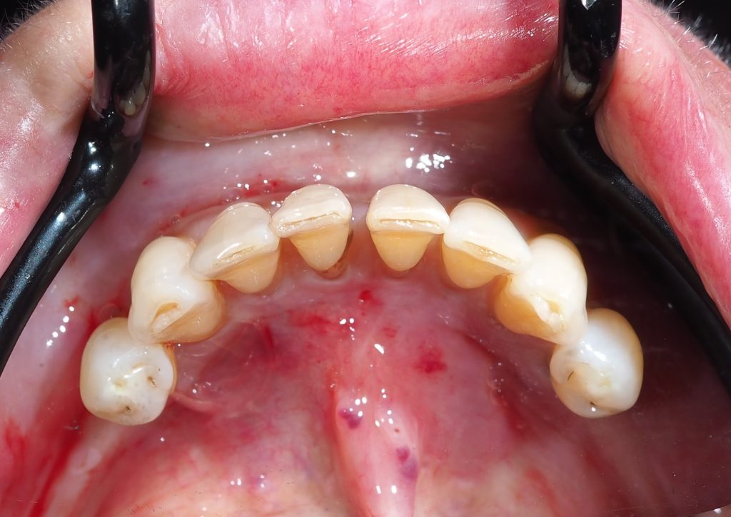
A 52-year-old female presented at FACE Dental in Coventry. Her main complaint was that she had a loose upper denture that rubbed against the gums and several of her lower teeth were mobile (Figures 1 and 2).
She was a non-smoker, did not suffer from bruxism and had a clear medical history. Upon assessment, the lower premolars were found to be grade 2 mobile, and the lower anterior teeth were grade 1 mobile. The patient had no temporomandibular joint clicking, and relatively good mouth opening and access. Occlusion was satisfactory and there were no contraindications for implant therapy. The patient’s ridge was 1 or 2 on the Cawood and Howell classification system for edentulous jaws. I therefore concluded that it would be adequate in height and width for implant placement.
Treatment Options
Alternatives to implants were initially discussed with the patient. The first option was to continue with the status quo. The second option would be to provide a better-fitting conventional upper denture and carry out periodontal treatment on the lower teeth. The patient’s third option would be to remove the mobile lower teeth and transition to new upper and lower conventional dentures.
Treatments including dental implants were also considered. The patient was offered the choice of bridges supported by implants on locators, or SKY fast & fixed with copaSKY implants to provide immediately loaded temporary bridges, transitioning to zirconia full-arch bridges at the end of treatment. Since the patient’s ridge was adequate, there was no need to consider pterygoid or zygomatic implants.
The patient expressed a strong preference for a fixed, full-arch option. The only choices available, therefore, were an implant-supported overdenture, or SKY fast & fixed with copaSKY implants. She opted for the latter as she did not want to wait or suffer the embarrassment of having no fixed teeth during the treatment period. We also wanted to achieve an outcome with the best possible success rate.
Initial planning and imaging
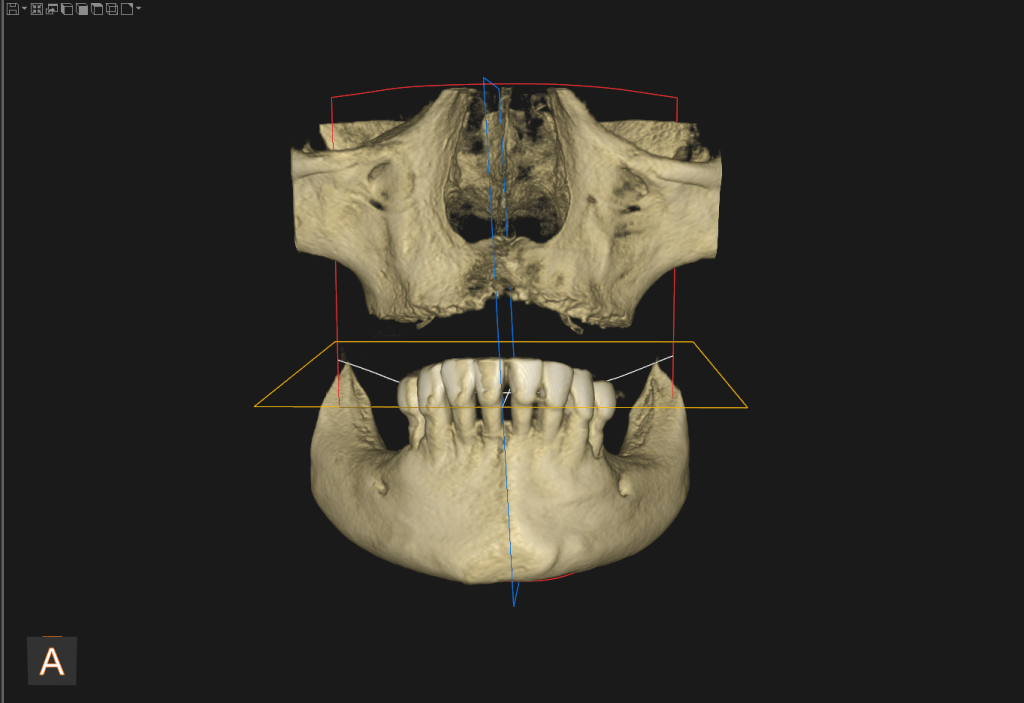
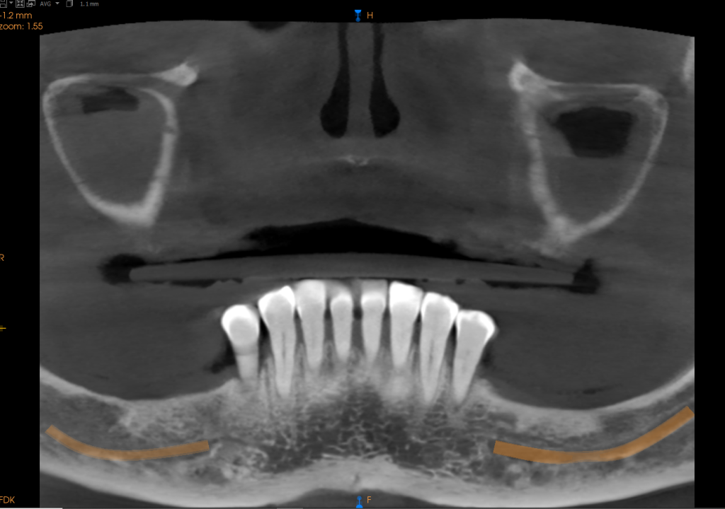
An intraoral scan was taken by our treatment coordinator, followed by extraoral photographs and a CT scan (Figures 3 and 4). They were sent to the laboratory for creation of the wax-up to enable me and the patient to visualise what her new implant-supported teeth would look like. In partnership with the laboratory, several discussions then took place with the patient about what would best suit her in terms of teeth size, shape and colour.
Before and after images were shown to the patient. Time and care were taken to assess the chosen position of her new teeth and check for adequate bone density and height in these areas. This was essential if we were to achieve a restoratively driven outcome, approaching the case with the endpoint in mind. Following the patient’s consent for the chosen treatment plan, we proceeded to the surgical phase.
I selected bredent medical copaSKY implants for this case as they have consistently provided me with high degrees of predictability and stability. The implant’s backtaper helps prevent bone loss and the narrow multi-units facilitate a much more aesthetic-looking prosthesis. The implants and SKY fast & fixed system, combined with accurate and time-saving photogrammetry, have benefitted our practice in so many ways. Adoption of photogrammetry in the planning of immediately loaded full-arch cases enables a fast, precise and efficient procedure, with good integration with other aspects of the digital workflow. The patient can be offered the very latest in cost-effective, innovative dental technology with speed of treatment, reduced waiting time, minimised risk of errors and complications, and a more streamlined pathway of care.
Same-day surgery
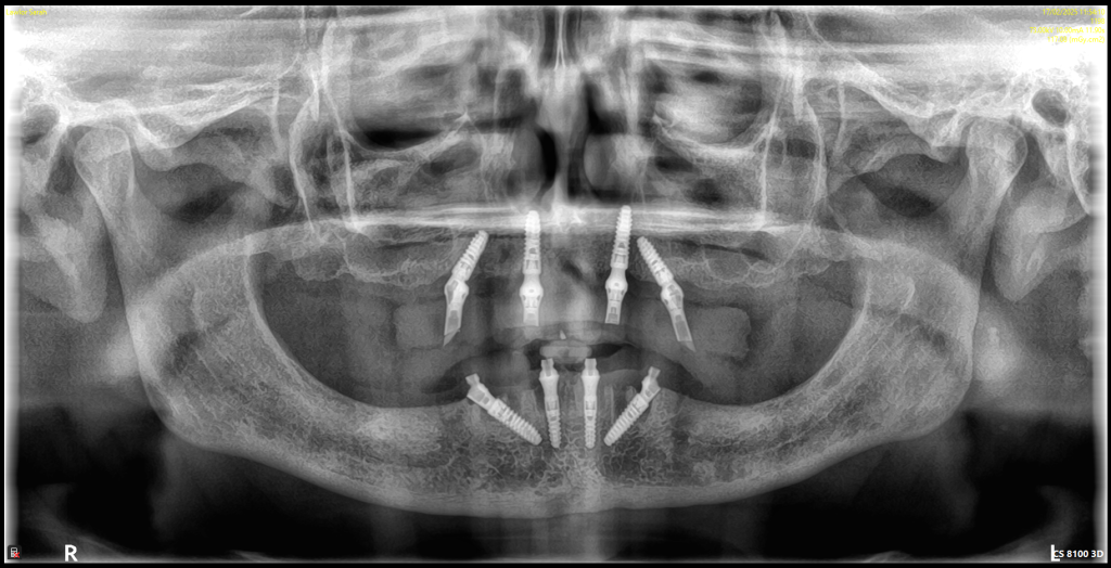
On the day of surgery, preoperative scans of the patient’s mouth were taken with an arch tracer. The arch tracer allows the laboratory to stitch all scans together after they are produced. The patient’s lower mobile teeth were extracted, the arch was exposed, the flap raised and four copaSKY implants placed subcrestally (Figures 5-7). Two 3mm by 10mm implants were placed in the anterior zone. Two 3.5mm by 12mm units were used for the angled implants. Placement was carried out with a restoratively driven approach, avoiding any critical anatomy. I used a prosthetic guide to show the final tooth position to help avoid excessive buccal placement.
The edentulous upper arch was then treated following the same method (Figures 8-11), with posterior placement of two 3.5mm by 10mm implants along the sinus wall and two 3.5mm by 12mm implants placed in the anterior zone. These needed to be longer, to anchor into the floor of the nose (Figures 12 and 13). All implants were placed at 45N-cm or higher, achieving a cumulative torque value of more than 200N-cm. My implant placement technique was freehand but in order to achieve high primary stability, particularly in the posterior maxilla, the osteotomy was under-prepared and a larger implant diameter was placed into each site.
Autogenous bone grafting, which in my experience gives the optimum healing response, was performed by harvesting material from the ridge reduction. After suturing with a simple interrupted technique, digital intraoral scans were taken with the bredent medical SKY uni.cone scan bodies on each multi-unit abutment (Figures 14-17). Photogrammetry scanning with the Tupel 3D machine was then carried out (Figure 18). The entire procedure is incredibly fast, taking around 15 seconds for the calibration and 15 to 20 seconds for scanning.
Bridge fabrication and restoration
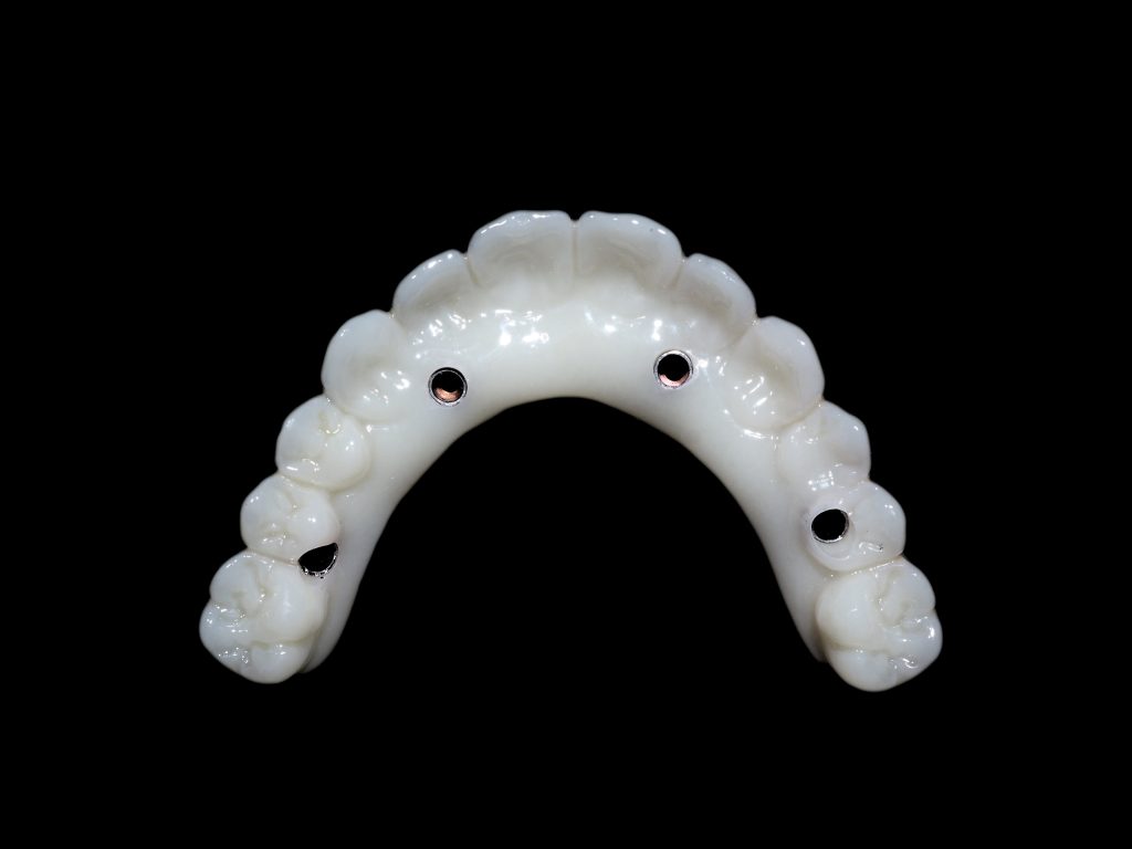
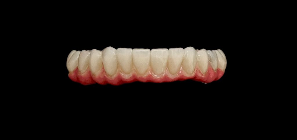
The laboratory’s preoperative wax-up was used to guide the location and design of the patient’s new teeth. The scans were imported into exocad, and the two prostheses were designed. The intraoral scans were stitched together with the photogrammetry scans and the wax-up was placed over them, allowing fabrication of the fully fixed, immediately loaded bridges (Figures 19 and 20).
The precision offered by the photogrammetry system acts like a jig for the full-arch bridge and reduces the steps in the restorative process. In this case, the passivity of the bridges was perfect. When the patient returned for the fitting later that day, the bridges were placed over the exposed multi-unit abutments and torqued down according to the manufacturer’s recommendations. The bridges were then sealed with polytetrafluoroethylene
(PTFE) and composite, and the patient was sent home to get accustomed to her new bite and interim prostheses. For the temporary bridges, our lead technician, Michael Joseph, selected G-CAM, a polymethyl methacrylate (PMMA) base reinforced with graphene, which helps to minimise the risk of breakages. This softer material would also enable the patient to get used to the prostheses.
The patient will return to us two months after surgery, when I will be able to adjust the prostheses and alter the occlusion if necessary. A total healing period of four months will be allowed, during which the patient’s expectations for function and aesthetics will be carefully managed. Following the healing period, the temporary bridges will be replaced with metal and zirconia frameworks.
Case Reflection
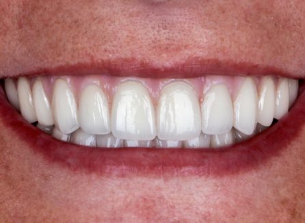
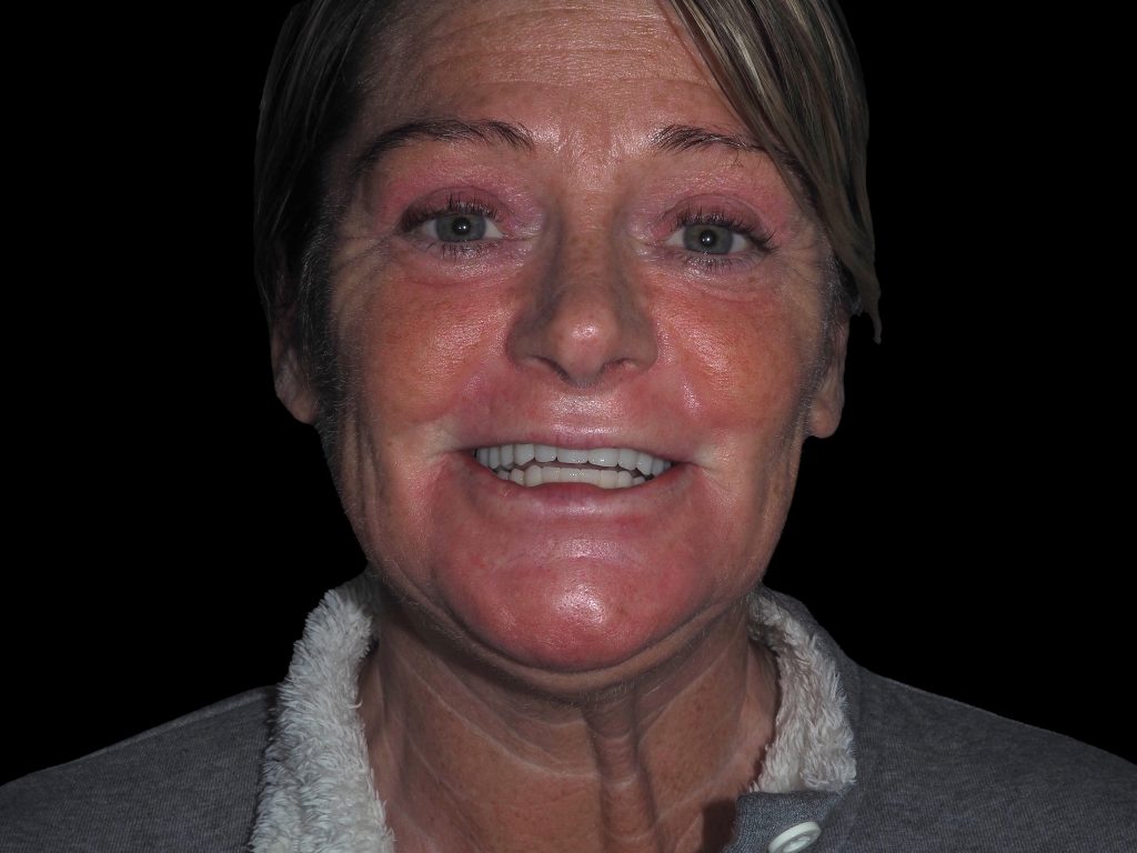
The treatment that was performed was truly life changing for this patient; this was evident in her eyes and her wide smile after we fitted the temporary bridges (Figures 21 and 22). She was able to leave the practice with her fixed teeth and smile restored in the most efficient and cost-effective way possible. I was very pleased with the result as well. The entire procedure went very smoothly, although I might consider offering sedation in the future, to further reduce any anxiety before treatment.
The only slight challenge of the case was the minimal bone in the upper left quadrant, in which I decided to place the implants following a palatal approach, with some thread exposure.
Planning for implant success
For any dental practitioner who is familiar with the art of immediate implants and bone grafting, full-arch teeth-in-a-day treatments are the next logical step of progression. Despite common myths, this doesn’t have to be a daunting prospect, with the right training and mentoring. The key is to base our approach on a restoratively driven outcome, working back from the desired end result. With meticulous planning, scanning and laboratory support we can improve predictability and reduce the risk of errors and complications. Once the procedure has been mastered, the same approach can be applied each and every time.
In addition to our choice of products, techniques and technology, it is also my belief that as a clinician, a healthy body and healthy mind are essential to complete the recipe for success. We are standing over patients for several hours during the surgical procedure and this requires core strength and stability. We also need to have a healthy state of mind to be in the best possible place to look after our patients at each step of the way.
About the author: Abdul Osman DMD MSc PGdip PGcert MInstLM
Dr Abdul Osman is the Principal Dentist and Director at FACE Dental and The Smile Centre in Coventry. He holds a postgraduate Certificate in Implantology, a postgraduate Diploma in Leadership and Management, and a Master’s degree in Dental Implants. He has also completed the world-renowned Advanced Surgical Master’s in Bone Regeneration with Professor Istvan Urban.
Abdul has a keen interest in providing patients with the most advanced dental implant treatments with digitally driven prosthetics and surgery. Dr Osman has lectured nationally and internationally for a number of prestigious bodies and was the implant programme lead at the College of Medicine and Dentistry in Birmingham. He now focuses his efforts on mentoring his community of young implant surgeons and running short courses on immediate implant placement. Abdul is currently the key opinion leader for a number of dental products including bredent’s SKY implants.
For further information:
FACE Dental, 76 Quinton Road, Coventry, CV3 5FD
Visit: www.facedental.co.uk
Tel: 024 7650 1125
Email: dr-ahosman@hotmail.com